
Art in Neuroscience 2021
Images from Cellular Imaging’s inaugural image and video competition showcasing some of the wonderful images produced by ZI staff, students, and affiliates.
-
Creative:
Awarded for the most creative and visually engaging science-related image. This can include images that have been digitally manipulated, image collages, renderings, and any images creatively exploring images from microscopy.
Winner receives a $150 gift card and framed print of their image.
Runner up receives $50 gift card.
Scientific:
Awarded for the most visually appealing scientific image that effectively conveys the research theme. The image should speak for itself, with digital manipulation limited to changes in brightness and contrast and coloration (e.g. selecting colors to differentiate individual fluorescent channels).
Winner receives a $150 gift card and framed print of their image.
Runner up receives $50 gift card.
Video Award:
Awarded by the judges to the most creative, interesting, or visually appealing video.
Winner receives a $150 gift card.
People’s Choice Award:
The most popular image as voted by the general public.
A framed picture will be awarded to the winning entrant and a voting member of the public.
-
Paula Croxson (Zuckerman Institute), Matt Woodward (Dear Mama), and Dister Rondon (I Love My Hood & ZI Art in Education Lab 2021 Resident)
Scientific Award | 1st Place

Salamander Senses
Eliza Jaeger | Tosches Lab
Staining performed with help from Alonso Ortega Gurrola
Funding: NSF GRFP
This is an image of an entire, cleared, larval salamander (Pleurodeles waltl) head with its brain and skin shown in white and developing sensory projections along its arms, skin, and gills shown in red.
Scientific Award | 2nd Place

BirdBow
Marissa Applegate, with help from Konstantin Gutnichenko | Aronov Lab
Funding: Beckman Young Investigators Award
Discovery of a possible entorhinal cortex analog in black-capped chickadees, revealed through anatomical tracing. Differently colored cells send input to different portions of the chickadee hippocampus.
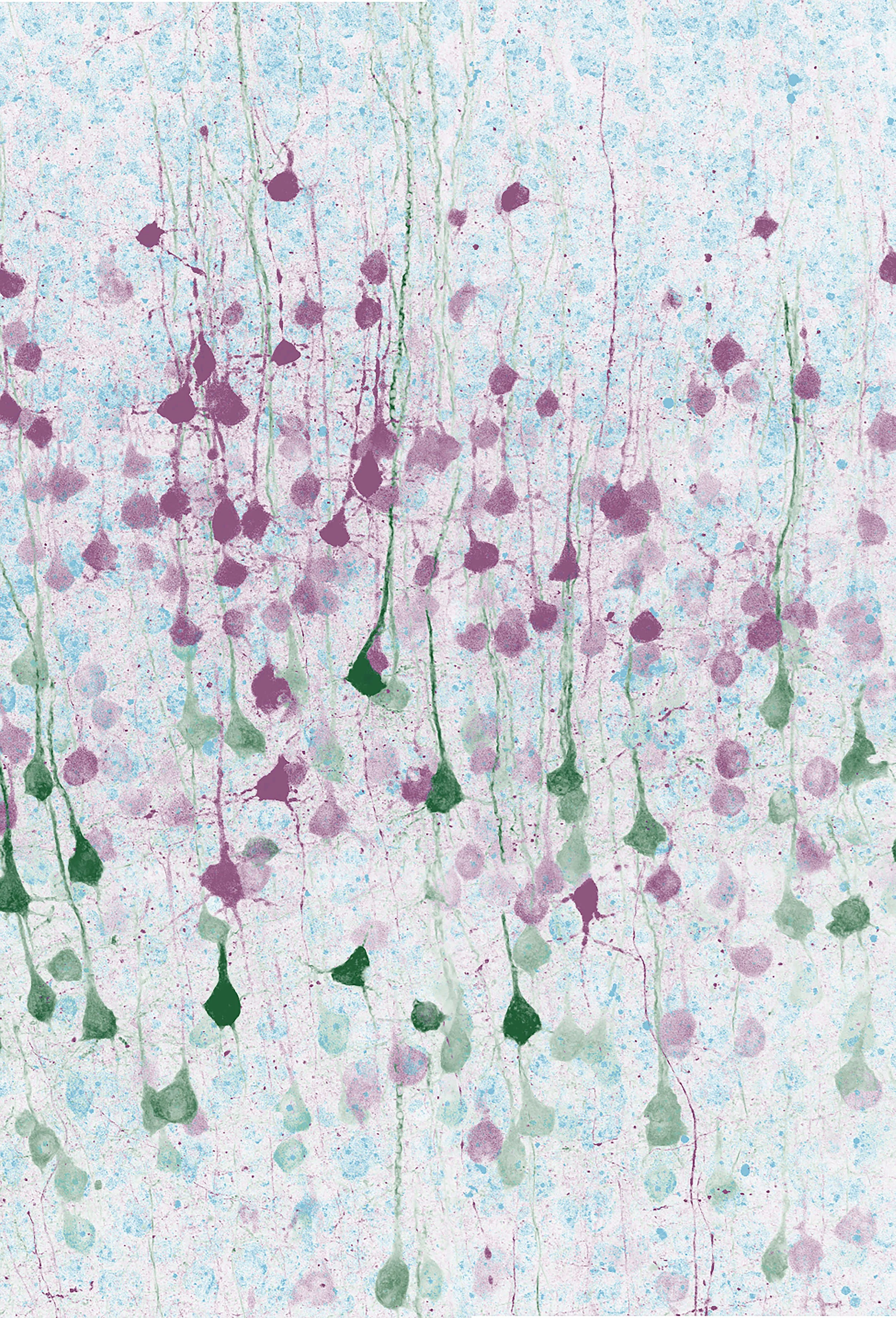
Creative Award | 1st Place

Spinal Inflorescence
Anders Nelson | Costa Lab
Funding: 1K99NS118053-01
Spinal interneurons expressing calbindin (purple) surrounded by excitatory synapses (pink).
Creative Award | 2nd Place

The perineuronal net extracellular matrix of the hippocampal CA2 subfield
Alex Whitebirch | Siegelbaum Lab
Funding: 1F31NS113466-01
The perineuronal net is a specialized extracellular matrix that typically ensheathes parvalbumin-expressing interneurons. However, in the CA2 subfield of the hippocampus this matrix is also found surrounding excitatory pyramidal neurons, where it has an important role regulating synaptic plasticity.
Peoples Choice Award

The sticky glue of our brain
Eugenie PEZE-HEIDSIECK (she/her) and Carlos DIAZ SALAZAR ALBELDA (he/him) | Polleux Lab
Funding: NOMIS and Fyssen Foundation
Glia comes from the Greek and means “glue”. We put all these cells, yet with very different roles and very different origins, in the same general basket of brain "glue". Microglia come from the immune system, they hunt down and “eat” damaged neurons or fight infections. They also play a crucial role in the elimination of synapses and therefore in the communication of neurons
Video Award
Lina Marcela Carmona | Costa lab
Funding: Helen Hay Whitney Fellowship
Movement dictates our interaction with the world, but all actions must first be learned. This process of motor learning involves many regions in the brain some of which are crucial as you begin to learn the skill and others which are involved as you start progressing towards proficiency. However, we do not have a global view of the total number of regions involved, the degree of their engagement, and their involvement as learning progresses. To examine this, mice were taught to pull a small wheel, a task which they can learn in a few weeks. By using the protein FOS, we can mark cells that are engaged as the mice learn this task. This video comprehensively shows the broad engagement of cells throughout the mouse brain during early motor learning.
Explore all of the inspiring image entries below.

ab>Intrahippocampal axonal projections of dorsal CA2 pyramidal neurons
Alex Whitebirch | Siegelbaum Lab
Funding: 1F31NS113466-01
CA2 pyramidal neurons form extensive axonal projections, seen here projecting throughout the intermediate hippocampus and establishing synaptic connections throughout the CA1, CA2, and CA3 subfields.
The perineuronal net extracellular matrix of the hippocampal CA2 subfield
Alex Whitebirch | Siegelbaum Lab
Funding: 1F31NS113466-01
The perineuronal net is a specialized extracellular matrix that typically ensheathes parvalbumin-expressing interneurons. However, in the CA2 subfield of the hippocampus this matrix is also found surrounding excitatory pyramidal neurons, where it has an important role regulating synaptic plasticity.

Neurodegeneration in the mouse hippocampus following pilocarpine-induced seizures
Alex Whitebirch | Siegelbaum Lab
Funding: 1F31NS113466-01
In order to model temporal lobe epilepsy, mice are administered the drug pilocarpine hydrochloride. This muscarinic acetylcholine receptor agonist induces seizures and a characteristic pattern of hippocampal neurodegeneration, in which CA2 pyramidal neurons and dentate gyrus granule cells (labeled with a stain against PCP4 in green) typically survive.

Convergence
Tristan Geiller | Losonczy Lab
Neurons have the unique property of interacting with one another through synapses. This image represents neurons distributed throughout the hippocampus (dark blue) that have synapses onto the same individual target (cyan).

Mauve Avenger
Anders Nelson | Costa Lab
Funding: 1K99NS118053-01
Corticostriatal neurons labeled with rabies (purple) surrounded by GABAergic inhibitory interneurons (green).
Spinal Inflorescence
Anders Nelson | Costa Lab
Funding: 1K99NS118053-01
Spinal interneurons expressing calbindin (purple) surrounded by excitatory synapses (pink).

Shadows of the Mind
Anders Nelson | Costa Lab
Funding: 1K99NS118053-01
Neurons in motor cortex expressing fluorescent reporters.
Mental Landscapes
Anders Nelson | Costa Lab
Funding: 1K99NS118053-01
Pyramidal neurons in a mouse motor cortex: corticospinal neurons (green); corticostriatal neurons (purple); cell nuclei are in light blue
Salamander Senses
Eliza Jaeger | Tosches Lab
Staining performed with help from Alonso Ortega Gurrola
Funding: NSF GRFP
This is an image of an entire, cleared, larval salamander (Pleurodeles waltl) head with its brain and skin shown in white and developing sensory projections along its arms, skin, and gills shown in red.

Amphibians Go Viral
Eliza Jaeger | Tosches Lab
Funding: NSF GRFP
This is a slice of an entire adult salamander (Pleurodeles waltl) brain with its brain cells shown in blue. The green cells have been injected with a virus called AAV PHP.eB expressing a fluorescent protein — a wonderful neuroscience
tool that hasn’t been used in amphibian brains before!

The brain clock’s protal system
Yifan Yao & Alana Taub | Silver Lab
Funding: NSF 1749500 & NIH 1S10OD023587-01
Blood vessels (green) from the brain clock suprachiasmatic nucleus (bottom left, magenta) reach a nearby highly vascularized region, the organum vasculosum of the lanima terminalis (right), forming a shortcut for diffusible signals between them.

Symphony of the Brain
Yow-Tyng (Tim) Yeh, with help from Qian Du | Woolley Lab
Labeling of the midbrain dopamine-producing cell population (red) and its subpopulation (green) that projects to the posterior striatum.

Maggot Map
Raffi Cohn and Samantha Shih | Grueber Lab
A Hemisegment of the Drosophila Larvae highlights the intricate and beautiful interwoven dendrites of different types of sensory neurons, including those that respond to pain (in red) and to touch (in green), with axons converging in the central nervous system on the right.

The stars within our brain
Eugenie PEZE-HEIDSIECK (she/her) | Polleux Lab
Funding: NOMIS and Fyssen Foundation
Long despised and set aside, astrocytes fulfill important functions of repair, regulation, maintenance of tissue, glutamatergic transmission, they also contact the blood capillaries with their “endfeet” ... the list is long and we cannot deny the beauty of their stary morphology.
The sticky glue of our brain
Eugenie PEZE-HEIDSIECK (she/her) and Carlos DIAZ SALAZAR ALBELDA (he/him) | Polleux Lab
Funding: NOMIS and Fyssen Foundation
Glia comes from the Greek and means “glue”. We put all these cells, yet with very different roles and very different origins, in the same general basket of brain "glue". Microglia come from the immune system, they hunt down and “eat” damaged neurons or fight infections. They also play a crucial role in the elimination of synapses and therefore in the communication of neurons.
BirdBow
Marissa Applegate, with help from Konstantin Gutnichenko | Aronov Lab
Funding: Beckman Young Investigators Award
Discovery of a possible entorhinal cortex analog in black-capped chickadees, revealed through anatomical tracing. Differently colored cells send input to different portions of the chickadee hippocampus.

This Feels Right
Kevin M. Cury | Axel Lab
Funding: Howard Hughes Medical Institute
Here we see the terminal abdomen of a female fruit fly. Sensory neurons (green) that connect to individual hairs are key to identifying soft places to lay eggs.

The World Within
Kevin M. Cury | Axel Lab
Funding: Howard Hughes Medical Institute
Here we see the reproductive system of a female fruit fly. Laying an egg requires the coordination of multiple muscle groups and organs.

Bitter neurons in amygdala
Li Wang | Zuker Lab
Funding: R01DA035025
The bitter neurons in the CEA were labeled in a Trap2-Ai9 mouse, stimulated by cycloheximide. The tSNE map shows single-cell clusters in the CEA (10x genomics).

Taste projections in mouse brain
Li Wang | Zuker Lab
Funding: R01DA035025
The view showing a CUBIC cleared mouse brain with projections from the sweet cortical field labeled with eGFP (GCsw, cyan) and the bitter cortical field labeled with tdTomato (GCbt, orange), published in Nature volume 558, pages 127–131 (2018).