
Art in Neuroscience 2023
Images from Cellular Imaging’s image and video competition showcasing some of the wonderful images produced by ZI staff, students, and affiliates.
-
Creative:
Awarded for the most creative and visually engaging science-related image. This can include images that have been digitally manipulated, image collages, renderings, and any images creatively exploring images from microscopy.
Winner receives a $150 gift card and framed print of their image.
Runner up receives $50 gift card.
Scientific:
Awarded for the most visually appealing scientific image that effectively conveys the research theme. The image should speak for itself, with digital manipulation limited to changes in brightness and contrast and coloration (e.g. selecting colors to differentiate individual fluorescent channels).
Winner receives a $150 gift card and framed print of their image.
Runner up receives $50 gift card.
Video Award:
Awarded by the judges to the most creative, interesting, or visually appealing video.
Winner receives a $150 gift card.
People’s Choice Award:
The most popular image as voted by the general public.
A framed picture will be awarded to the winning entrant and a voting member of the public.
-
Melissa Becker | VP Business Development (Manhattanville Market)
Ben Dubin-Thaler, Ph.D. | Founder & Executive Director - BioBus
Art in Neuroscience 2023
Scientific Award | 1st Place

Envisioning Eye Development
Margarita Dillinger, she/her | Mason Lab
Funding: NIH R01 EY015290, an António Champalimaud Vision Award, Simons Foundation Senior Fellow Award, and the National Organization for Albinism and Hypopigmentation (NOAH)
Embryonic mouse eye immunostained for DNA (blue), a regulator of cell division (CyclinD2) (red), and Zic2, transcription factor in ipsilateral retinal ganglion cells (green)..
A touching image of a Drosophila larva
Wes Grueber, submitted on behalf of Dr. Samantha Galindo | Grueber Lab
Funding: NINDS of the NIH Award Number R01NS061908 National Institutes of Health NS098765
Image of a Drosophila larva with nociceptive (noxious sensing) neurons in green and touch sensing neurons in magenta. Both groups of somatosensory neurons reside just inside a transparent body wall, which they cover completely without gaps or overlap.

Scientific Award | 2nd Place
Creative Award | 1st Place

Venetian Mask
Panagiota (Julie) Apostolou | Gogos Lab
A part of the brain that is mainly responsible for memory, called the hippocampus fixed at the time that some of the neurons become activated.
Creative Award | 2nd Place
Where vision meets movement
Stephen Huston | Huston Lab
An image of a fly brain (grey) showing brain cells that respond to visual motion (magenta) connecting to a motor neuron (green) that controls the fly’s muscles. Studying these simple neural circuits that translate sensation to movement helps us understand how the brain generates behavior. Image made in collaboration with Rebecca Johnston, Friday Harbor Laboratories. .

Peoples Choice Award
Venetian Mask
Panagiota (Julie) Apostolou | Gogos Lab
A part of the brain that is mainly responsible for memory, called the hippocampus fixed at the time that some of the neurons become activated.
Explore all of the inspiring image entries below.

Jackson Pollock’s drips and synaptic FLPS
Laudan Nikoobakht & Alana Mendelsohn | Costa Lab
Green cells are cells expressing Pou6f2 (gene). Red and yellow neurons were ‘flipped’ using a FLP recombinase to reveal which Pou6f2 neurons have GABAergic synapses.

Spoderman into the Neuroverse
Laudan Nikoobakht & Zirong Gu | Costa Lab
The brain of a mouse bit by a radioactive spider (In other words: injection site of parafascicular nucleus of the thalamus injected with a retro AAV. Retrograde tracers outline neurons from the terminals of their axons to their cell bodies.)

Dual purpose brain
Ines Rodrigues-Vaz | Costa/Peterka Labs
Funding: FCT Portugal PD/BD/105950/2014; Simons-Emory; International Consortium (Simons 717104); U19 NS104649 (to RC); ASAP-020551
Using genetics to see neural activity, and virus to induce neural activity.

Columns and bucket of neurons
Ines Rodrigues-Vaz | Costa/Peterka Labs
Funding: FCT Portugal PD/BD/105950/2014; Simons-Emory; International Consortium (Simons 717104); U19 NS104649 (to RC); ASAP-020551
Active neurons in the brain – from cortical columns to messy striatum.

Paint by Glia
Davys Lopez (he/him) | Mann Lab
Funding: R01NS070644
The colors highlight the wrapping glia of the immature fruit fly nervous system that facilitate neuronal communication to the muscles of the larval body.

Keeping a level head
Stephen Huston | Huston Lab
An image of the cuticle deep within a fly’s neck. When the fly rotates its head, tiny hairs (false colored red) are squashed by a flap of cuticle (false colored transparent blue). Each hair is monitored by its own sensory neuron which conveys information to the fly’s brain about its head posture. Image made in collaboration with Igor Siwanowicz, Janelia Research Campus.
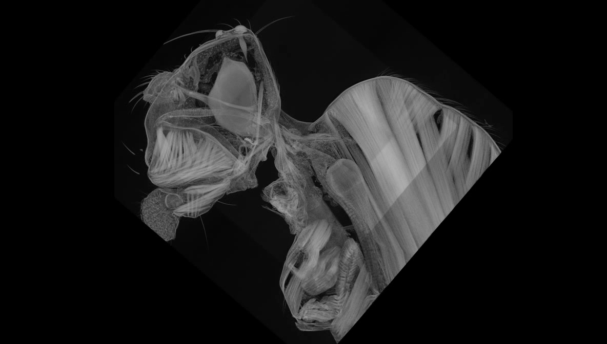
Fly muscles
Stephen Huston | Huston Lab
A micro-CT image of a fly’s head and upper thorax showing its muscles and brain. Image made in collaboration with Nirmala Iyer, Janelia Research Campus.

Where vision meets movement
Stephen Huston | Huston Lab
An image of a fly brain (grey) showing brain cells that respond to visual motion (magenta) connecting to a motor neuron (green) that controls the fly’s muscles. Studying these simple neural circuits that translate sensation to movement helps us understand how the brain generates behavior. Image made in collaboration with Rebecca Johnston, Friday Harbor Laboratories.

Venetian Mask
Panagiota (Julie) Apostolou | Gogos Lab
A part of the brain that is mainly responsible for memory, called the hippocampus fixed at the time that some of the neurons become activated.

409981 pictures of an HIV envelope protein
Nicholas C Morano | Shapiro Lab
Funding: Vaccine Research Center, National Institution Health
A 3D image reconstruction of 409981 electron microscopy pictures of an antibody (blue and red) bound to the HIV Envelope protein (gray and magenta) from a collaborative effort between the Shapiro Lab and the Vaccine Research center aimed at developing an HIV vaccine.

Envisioning Eye Development
Margarita Dillinger, she/her | Mason Lab
Funding: NIH R01 EY015290, an António Champalimaud Vision Award, Simons Foundation Senior Fellow Award, and the National Organization for Albinism and Hypopigmentation (NOAH)
Embryonic mouse eye immunostained for DNA (blue), a regulator of cell division (CyclinD2) (red), and Zic2, transcription factor in ipsilateral retinal ganglion cells (green).

A touching image of a Drosophila larva
Wes Grueber, submitted on behalf of Dr. Samantha Galindo | Grueber Lab
Funding: NINDS of the NIH Award Number R01NS061908 National Institutes of Health NS098765
Image of a Drosophila larva with nociceptive (noxious sensing) neurons in green and touch sensing neurons in magenta. Both groups of somatosensory neurons reside just inside a transparent body wall, which they cover completely without gaps or overlap.